


Martin Lardy


Pierre-François Lenne s’intéresse à la gastrulation, une étape essentielle dans la création des premiers tissus.
Et pourtant ils diffusent!
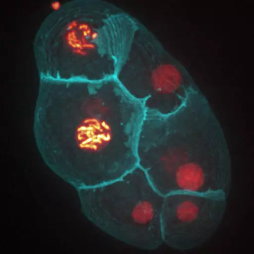
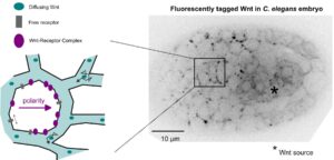
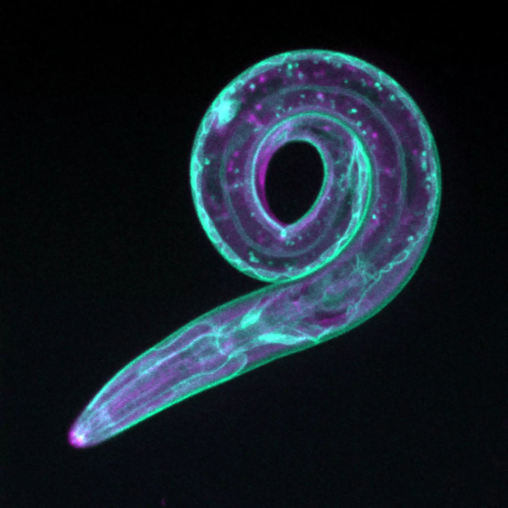
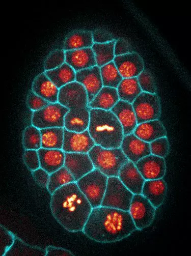
Dans les embryons de C. elegans les ligands Wnt diffusent dans le tissu pour polariser des cellules à distance.
Lost in translation: Vangl2 short and long
La traduction commence par une Méthionine: Vrai, mais pas toujours, comme le montre une étude sur Vangl2.
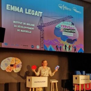
Le cerveau : un chantier permanent
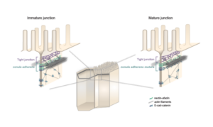
Les équipes Lenne et Le Bivic démontrent une nouvelle organisation pour les jonctions d’adhérence intestinales.
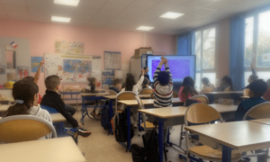
L’IBDM inspire les jeunes esprits : en impliquant les enfants des écoles primaires dans la lutte contre le cancer pédiatrique (“Contre le cancer, j’apporte ma pierre”) et en interagissant avec les lycéens grâce à des expériences immersives (DECLICS).
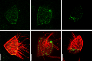
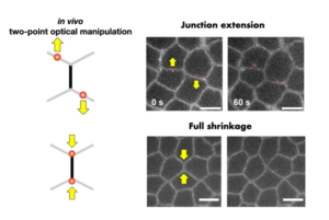
Les tissus épithéliaux sous tension: une étude explore comment les cellules se déforment individuellement et résistent aux forces.
Le groupe Lenne, ainsi que trois autres groupes, Merkel (CNRS), Trivedi (EMBL) et Ruprecht (CRG), se lancent dans ce projet !
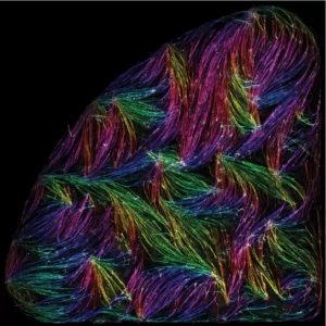
Les cellules musculaires s’auto-organisent en faisceaux de fibres in vitro, sans la présence de signaux externes !
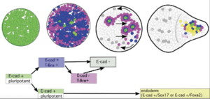
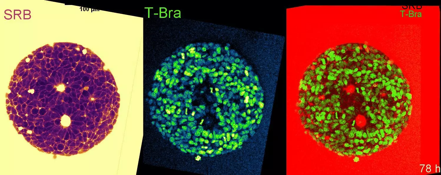
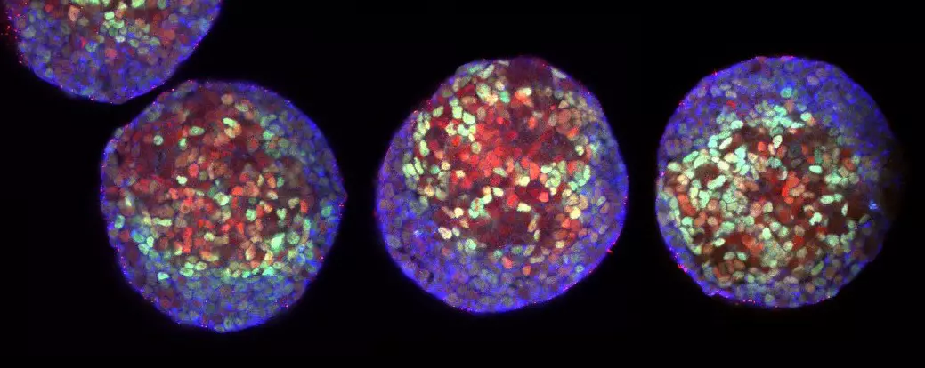
Cell-state transitions and collective cell movement generate an endoderm-like region in gastruloids
L’équipe de Lenne a publié dans Elife : En utilisant des gastruloïdes (agrégats 3D de cellules souches embryonnaires de souris), l’équipe a étudié, à une résolution cellulaire, la spécification de l’endoderme.
7 équipes de l’IBDM ont reçu une bourse ANR
7 équipes de l’IBDM ont reçu des subventions de l’Agence Nationale pour la Recherche (ANR) en 2021. Félicitations à Vincent Bertrand, Harold Cremer, Pascale Durbec,
Pierre-Francois Lenne élu membre de l’EMBO
Le 7 juillet 2020, 63 scientifiques de renom ont été élus membre de l’European Molecular Biology Organization (EMBO), en reconnaissance de leurs remarquables réalisations dans le domaine des sciences de la vie.
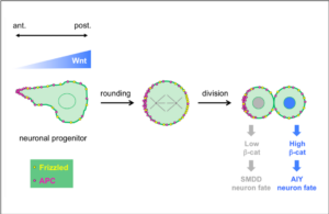
Dans une étude collaborative publiée dans Development, les équipes de Vincent Bertrand et de Pierre-François Lenne ont analysé le rôle de ligands Wnt lors des divisions générant des neurones au cours du développement du système nerveux.
Dans une étude récente parue dans la revue internationale Nature, Thomas Lecuit et ses collègues de l’Institut de Biologie du Développement de Marseille décrivent comment les changements de forme des tissus sont auto-organisés.