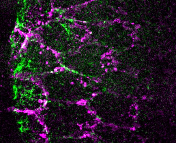
During embryonic development tissues undergo complex choreographies of morphological shape changes to form fully functional organisms, a process known as morphogenesis. Active forces, often due to cell contractility and adhesion, power these shape changes. How these forces are orchestrated in space and time to properly shape tissues is poorly understood. In this study recently published in Developmental Cell, Claudio Collinet and Anaïs Bailles in the group of Thomas Lecuit, focused on a wave of actomyosin contractility and tissue invagination, that drives the embryonic body axis extension of the fruit fly Drosophila, to understand how genetic patterning of mechanical cues and the shape of the tissue impact on morphogenesis. The authors zoomed on the process using live imaging microscopy and discovered that that the wave of tissue invagination emerges from an orchestrated sequence of interaction with the extraembryonic membrane enveloping the embryo called the vitelline membrane. Cells adhere to this envelope, activate MyoII and de-adhere from it in a wave. Collinet et al. identify two mechanochemical feedback processes that regulate cell contractility. They also uncover how the geometric configuration of the tissue impacts on the mechanical forces involved in tissue invagination. This study illustrates how mechanical and geometrical feedback mechanisms regulate tissue morphogenesis.
IBDM Portraits – Ahmed Fatmi
For me, building a qPCR technical platform is much more than a technical achievement.


