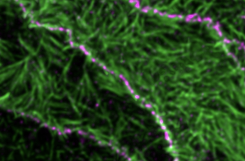
Pierre Mangeol together with the Lenne and Le Bivic labs used STED super-resolution microscopy to decipher how polarity proteins, the main organizers of epithelial cells, are themselves organized. This work led to two surprising findings. First, all these proteins organize as clusters of 100 to 200 nm. Second, while the literature predicted that a wide combination of proteins could interact with each other, making polarity very difficult to apprehend, the observations show that only two of these combinations of proteins are prominent. This establishment of a hierarchy in polarity protein interactions will help tackle the very complex cell polarity field with a new perspective.
To know more
Contact
Figure legend
The polarity protein PALS1 (magenta) at the apical junction of epithelial cells and actin (green) in microvilli


