Team members

Cellular Interactions, Neurodegeneration and Neuroplasticity

We investigate the organization and functional dynamics of brain neuronal circuits, their adaptive capacities and the mechanisms underlying their pathological dysfunction and neurodegeneration, with focus onto the basal ganglia network.

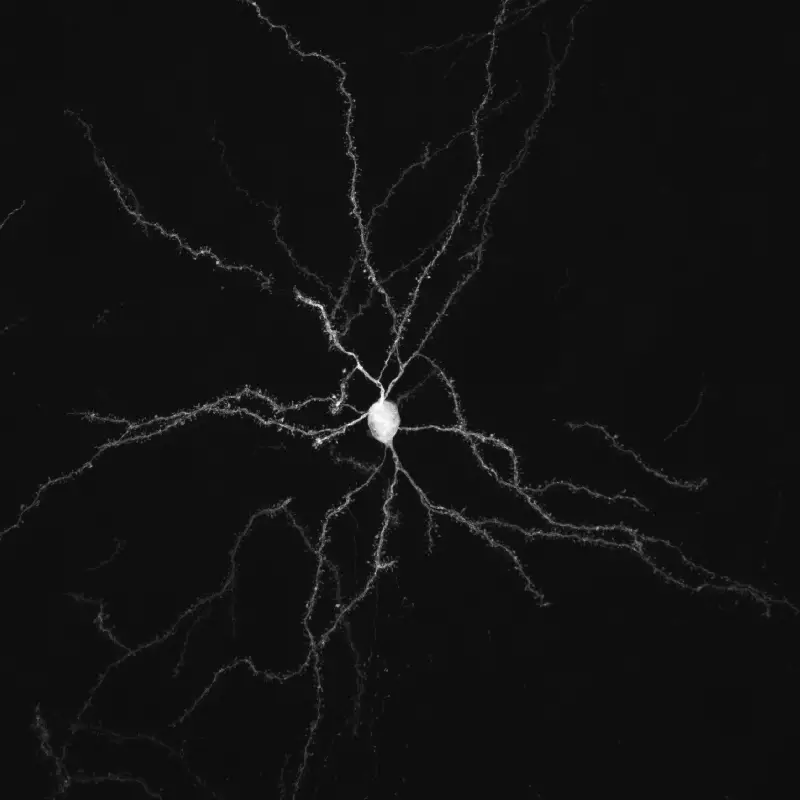
Our main research interest is on synaptic transmission, neurodegeneration and neuroplasticity in the adult brain. Communication between neurons at the level of their connections, called synapses, is the substrate of information processing in the networks underlying brain functions. Synaptic transmission is a highly dynamic and regulated process, influenced by glial cells, whose abnormalities are associated with a number of brain diseases (concept of synaptopathies). Neurodegeneration is a pathological process that triggers progressive dysfunction and death of nerve cells. Understanding the pathological mechanisms triggering and sustaining neurodegeneration (pathogenesis), its consequences on  circuit function and the mechanisms that help neurons to manage cellular stress is essential for the development of curative or disease-modifying treatments for devastating neurodegenerative disorders, such as Parkinson’s disease (PD). Neuroplasticity refers to the capacity of the nervous system to adapt in response to experience and to internal or external stimulations by modifying the interactions between nerve cells, including changes in the number of synapses and the efficacy/strength of synaptic transmission (synaptic plasticity), or by generating new nerve cells. This faculty is not limited to development, but occurs lifelong, although declining with aging. Neuroplasticity has been notably involved in learning and memory processes. It also occurs under pathological conditions or in response to chronic treatments. Such adaptive changes can represent compensatory mechanisms counteracting the deficits triggered by neuronal dysfunction or death, delaying the symptom onset, or, on the contrary, can participate in or even aggravate the deficits.

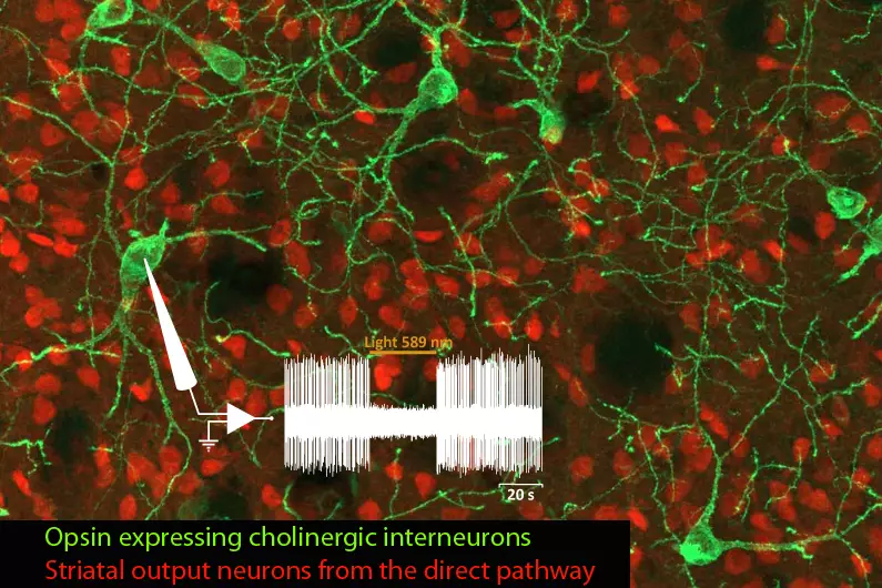
The team is investigating these processes in the context of basal ganglia (BG)-related functions and pathologies, in particular PD, a movement disorder characterized by the degeneration of midbrain dopamine neurons innervating the striatum, the main BG input station. Through collaborations, our work also addresses fundamental and clinically-relevant issues in the context of other neuropathologies, including autism spectrum disorder (ASD), Alzheimer’s disease and Charcot-Marie-Tooth disease.

Golgi staining-like view of a striatal medium-sized spiny neuron obtained by viral retrograde tracing, which reveals its thin dendritic arborizations and high density of spines that are primary post-synaptic seats of excitatory connections.

Publications

Our last publications

News

of the teams

Team members

They drive our research

Alumni

They contributed to our research
Maxime Assous
Research Associate, Center for Molecular and Behavioral Neuroscience, Rutger University, Newark, NJ, USA
Abid Oueslati
Associate Professor (Department of Molecular Medicine, Laval University), Director of the Molecular and Cellular Neurodegeneration Laboratory, CHU Research Center, Quebec City, Canada

Funding bodies

They support our research
ARN
Fondation Alzheimer
Fondation de France

Gallery

Cellular Interactions, Neurodegeneration and Neuroplasticity

The team investigates synaptic transmission, neurodegeneration and neuroplasticity in the context of BG-related functions and pathologies, with a focus on PD. Our approach combines the use and development of relevant experimental models in rodents (chronic deep brain stimulation, cell specific ablation, optogenetic or chemogenetic modulation of neuronal activity) with a variety of analytic tools, including behavioral tests, functional anatomy, tract-tracing and electrophysiology and is implemented with genetics, imaging and molecular biology in drosophila models. Ongoing research lines have two main objectives:

1. Gain knowledge on the anatomofunctional organization of the BG network and its remodeling in pathological condition, which may help design novel symptomatic treatments.

Our current work is centered on striatal cholinergic interneurons (CINs) and on corticostriatal synaptic function/plasticity in two pathological conditions: PD and ASD.

Regarding PD, we previously provided evidence for a causal role of CINs in parkinsonian symptomatology by showing that their optogenetic inhibition alleviates PD-like motor deficits (Maurice et al., 2015; Ztaou et al., 2016). Ongoing work aims at providing mechanistic insights into the antiparkinsonian potential of targeting these interneurons. We are challenging the hypothesis that abnormalities in CINs signaling contribute to altered corticostriatal transmission and plasticity in PD state, and that this implication depends on the thalamostriatal information they integrate. Our most recent data show that inhibiting CINs potentiates corticostriatal transmission in D1 receptor-expressing projection neurons, partially restores plasticity at corticostriatal synapses and alleviates motor-skill learning deficits in a mouse model of PD (Laverne et al., 2022).

Regarding ASD, we are pursuing a collaborative work with the team of Laurent Fasano at the IBDM that associated heterozygous deletion of TSHZ/Tshz3 with ASD (Caubit et al., 2016), a neurodevelopmental disorder defined by social interaction deficits and repetitive behaviors. The characterization of mouse models of conditional Tshz3 deletion from cell to behavior further showed that TSHZ3 plays a crucial postnatal role in the functioning of the corticostriatal circuitry (Chabbert et al., 2019; Caubit et al., 2021), and that dysfunction in cortical projection neurons or in CINs segregates with distinct core features of ASD, respectively social interaction deficits and repetitive/stereotyped behavior (Caubit et al., 2022). Ongoing work aims at deciphering the mechanisms of CIN dysfunction, its consequences on the functioning of the corticostriatal circuit, as well as the links with the striosome/matrix organization of the striatum and stereotyped behaviors.
Double labeling, in the striatum of transgenic mice, of the striatal output neurons expressing the dopamine D1 receptor (red fluorescence) and the cholinergic interneurons (green fluorescence) in which halorhodopsin expression allows their photo-inhibition. The electrophysiological trace illustrates the interruption of the spontaneous spiking of one recorded cholinergic interneuron when amber light is delivered in vivo into the striatum.â’¸ Nicolas Maurice, IBDM, Marseille.

2. Identify players in neuron death or defense pathways against cellular stress that may represent new targets for disease-modifying strategies.

This research is performed in the context of PD, but also of Alzheimer’s disease and Charcot-Marie-Tooth disease.

A first part is centered on the mechanisms regulating autophagy, mitochondrial dynamics and mitochondrial quality control via mitophagy, which are main players in neurodegenerative diseases. We are pursuing the study of the role in the brain of tumor protein 53-induced nuclear protein 1 (TP53INP1), a stress-induced protein known to act as a dual regulator of transcription and of autophagy, whose deficiency has been linked to cancer and metabolic syndrome through mechanisms that are also involved in neurodegenerative diseases (including chronic inflammation, oxidative stress and autophagy dysregulation). In collaboration with the groups of Alice Carrier (CRCM, Marseille) and Olga Corti (ICM, Paris), we recently uncovered a neuroprotective role of TP53INP1 for dopamine neurons under aging and PD-related conditions and provided evidence that TP53INP1 might maintain neuronal homeostasis by adapting basal mitophagy demands via autophagy regulation (Dinh et al., 2021). Ongoing experiments investigate the impact of TP53INP1 deficiency in the context of aging- and AD-related cognitive impairments, in collaboration with the team of Pascale Durbec at the IBDM. We are also interested in mitochondrial dynamics, whose alteration has been implicated in neurodegenerative disorders and hereditary neuropathies. In particular, we are studying the consequences of mitochondrial fusion and fission imbalance in drosophila models of Charcot-Marie-Tooth neuropathy type 2A through a variety of in vivo approaches.

Mitochondrial network in living larval drosophila motor neurons
A rare observation of drosophila mitochondria staring into the eyes of the experimenter. â’¸ Thomas Rival, IBDM

The second part aims at revisiting the involvement of the glutamate system in PD pathogenesis and pathophysiology. We previously developed a rat model mimicking the slowly progressing and asymmetrical degenerative process that characterize PD (Assous at al., 2014). In collaboration with the group of Franck Durif (University of Clermont Auvergne, CHU, CNRS), we are investigating the interhemispheric reactive changes affecting the main BG glutamate components in this model and their possible contribution to the evolution of nigrostriatal dopamine neuron death, using a combination of in vivo and ex-vivo approaches.

Progressive loss of dopamine neurons in the substantia nigra pars compacta (SNc delineated by dotted lines) in a model of PD based on dysfunction of excitatory amino acid transporters.

In parallel to these main research lines, we are involved in a collaborative project developed by Rosanna Dono in the team of Flavio Maina at the IBDM, which aims at improving the efficacy and safety of the cell-based replacement therapy for PD using human induced pluripotent stem cells (hiPSCs). The objective is to evaluate the potential of regulating the levels of glypican 4, a signaling modulator, as a strategy to foster hiPSCs differentiation towards the disease-relevant cell type, namely ventral midbrain dopamine neuron, and reduce their propensity to develop tumors upon brain transplantation in a rat PD model at different stages of in vitro differentiation (Corti et al., 2021).