Les différents types de muscle peuvent produire des forces endurantes moyennes ou des forces maximales élevées, suivies d’une fatigue rapide. L’énergie alimentant leurs moteurs contractiles est en grande partie fournie par leurs mitochondries. En combinant la génétique chez la drosophile avec l’imagerie de pointe et le “deep-learning”, des chercheurs de l’Institut de biologie du développement de Marseille ont découvert que les mitochondries coordonnent leur formation avec le développement des myofibrilles pour correspondre au bon type musculaire. Pour ce faire, les mitochondries et les myofibrilles subissent des interactions physiques hautement orchestrées qui donnent des instructions sur la façon dont chacune d’entre elles progresse au cours du développement. Ces résultats montrent que la morphogenèse des mitochondries et des myofibrilles est liée mécaniquement pour définir le destin correct du muscle. Ces résultats sont maintenant publiés dans Nature Communications.
– Plus d’informations en anglais –
Most animals have different muscle types dedicated for specific tasks. These can twitch slowly and involuntary, such as the ones that ensheath our gut or keep our heart beating, or be fast and quickly exhausted, like the ones we use to generate maximum force for escaping or lifting heavy objects. Mitochondria are the power houses of all muscles and generate most of their energy by converting nutrients from food into ATP, which fuels the muscle motor proteins. Mitochondria adopt different shapes and localisations in different cell types, in particular in different muscle types. How this is achieved is largely unknown. Now, a multidisciplinary project led by Nuno Luis and Frank Schnorrer at the Developmental Biology Institute of Marseille (IBDM) of the CNRS & Aix-Marseille University, has found that the force producing myofibrils coordinate their development closely with their mitochondria by generating a mechanical feedback mechanism that instructs their final morphology and function.
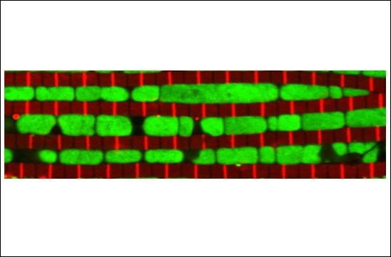
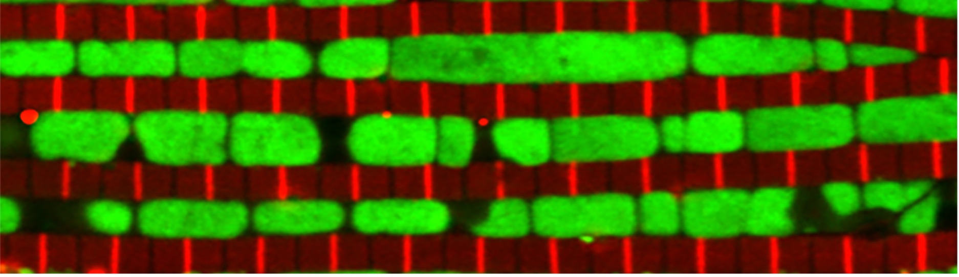
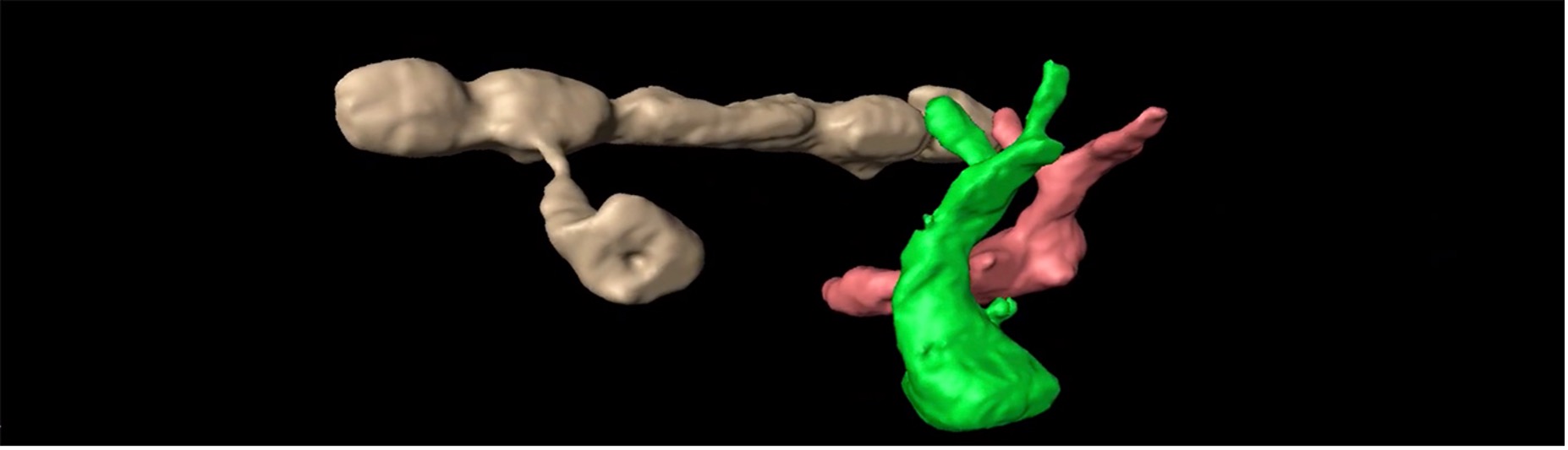
The flight muscle of the fruit fly Drosophila melanogaster is a “stretch-activated” fibrillar muscle type, similar to the mammalian heart. One group of muscles mechanically senses when stretched by the other group, and then contracts. Hence, wings can oscillate 200 times per second to enable flight. These oscillations require an extensive amount of ATP. Thus, the mitochondria are located in intimate contact to and squeezed in between the individual myofibrils, to quickly deliver ATP to the motor proteins. In contrast, the slower leg muscles of the fly used for walking display a different mitochondrial morphology by forming complex shapes above or below the myofibril layer.
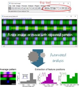
By using state-of-the-art 3D imaging and deep-learning algorithms, Jerome Avellaneda, PhD student and first author of this study, discovered that the muscle-type specific mitochondrial morphology is already instructed during the early stages of flight muscle development. Initially, mitochondria are clustered together. However, as soon as the myofibrils assemble mitochondria intercalate in between them to insulate one from its neighbours. In the following steps, myofibrils grow in size and diameter and push their surrounding mitochondria into elongated ellipsoid shapes. The authors found that the mitochondrial fusion and fission machinery is crucial for this dynamic “mitochondrial-myofibril-dance” with fission being necessary for the mitochondria to intercalate between the myofibrils. Surprisingly, a failure in intercalation results in a myofibril organisation resembling the ones in the slow leg muscle. Even more surprisingly, these flight muscles express leg muscle specific contractile proteins suggesting that the mitochondrial morphology directly feeds back on muscle type choice. These results suggest that in force producing muscle tissue the morphogenesis of the different cellular components and organelles is highly coordinated. Interfering with one, may dramatically change the other. This may have important consequences for treatment of muscle and in particular heart disease in human.
En savoir plus :
Contact
Frank Schnorrer – frank.SCHNORRER@univ-amu.fr