Post en anglais
Researchers have used the CERN pixels to follow tumour evolution in a liver cancer mouse model.
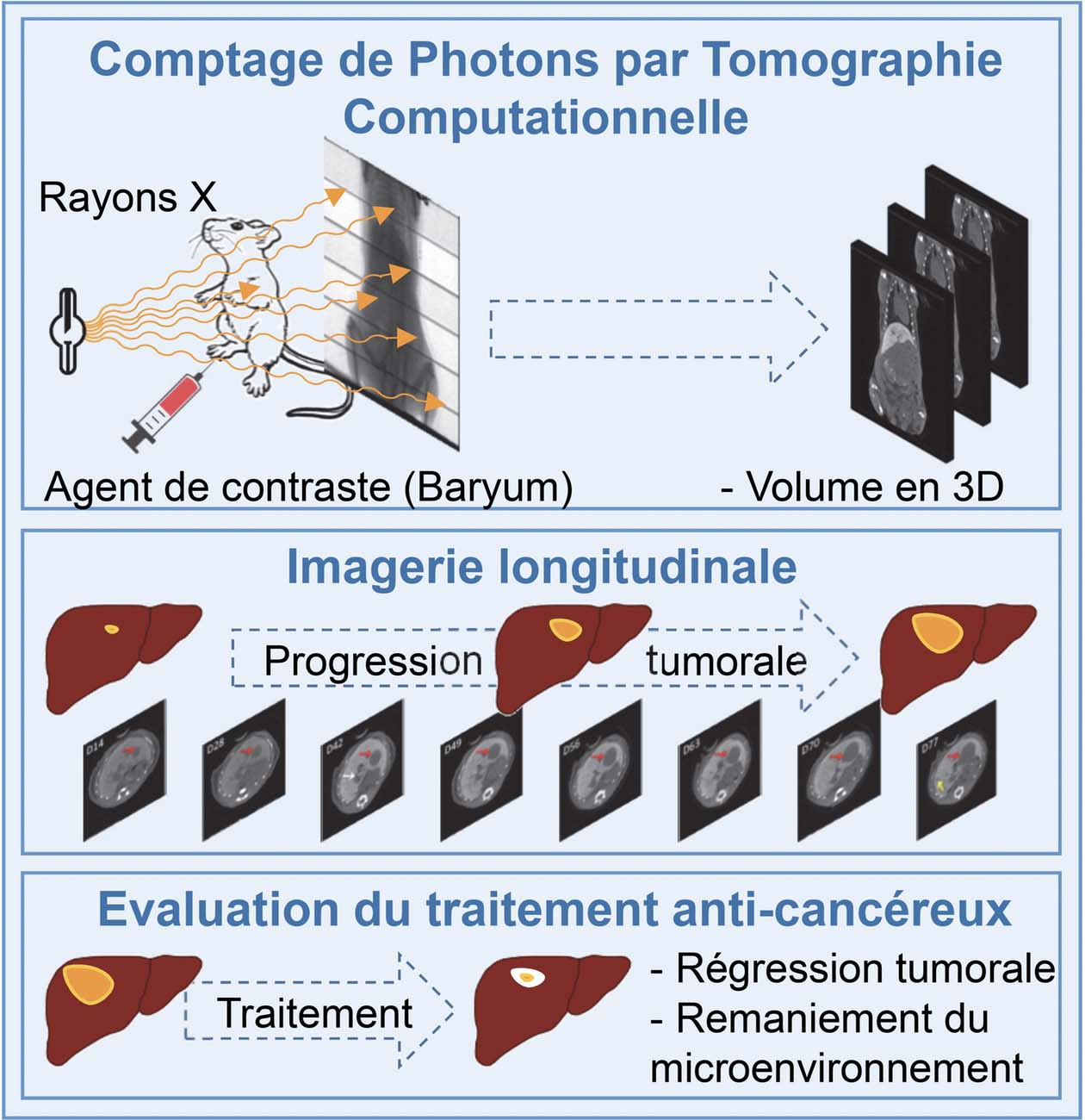
A photon counting scanner has been developed by engineers and physicists at the CPPM for longitudinal in vivo imaging studies with Maina Team to follow for several months the tumour growth of mice spontaneously developing liver tumours. We found a remarkable heterogeneity in the dynamics for tumours at the initiation phases, whereas the growth curve of evolving tumours exhibited a comparable exponential growth, with a constant doubling time. Furthermore, this PC-CT imaging system demonstrated the effectiveness of a combinatorial therapy leading to a drastic tumour regression accompanied by a striking remodelling of macrophages in the tumour microenvironment. This interdisciplinary study was published in the journal iScience.
To know more :
Loriane Portal1,#, Franca Cassol1,#, Sylvie Richelme2, Mathieu Dupont1, Yannick Boursier1, Ahmed Abdouni2, Nathalie Auphan3, Lionel Chasson3, Samantha Fernandez4, Laure Balasse4, Rosanna Dono2, Fabienne Lamballe2, Benjamin Guillet4, Toby Lawrence3, Christian Morel1,*, Flavio Maina2,*
1Aix-Marseille Univ, CNRS/IN2P3, CPPM, Marseille, France
2Aix-Marseille Univ, CNRS, IBDM, Marseille, France
3Aix-Marseille Univ, CIML Marseille, France
4Aix-Marseille Univ, CERIMED, Marseille France
Summary: Computed Tomography is a powerful medical imaging modality for longitudinal studies in cancer to follow neoplasia progression and evaluate anticancer therapies. We have generated a Photon Counting micro-Computed Tomography (PC-CT) method based on hybrid pixel detector with enhanced sensitivity and precision of tumour imaging. Additionally, the lower X-ray dose radiation required for PC-CT allowed multiple imaging up to 3 months. We then applied PC-CT for longitudinal imaging in a clinically relevant liver cancer model, the Alb-R26Met mice, which offers the unique possibility to follow initiation, latency, and evolution of spontaneous liver tumours, in contrast to other preclinical systems based on single tumour formation following experimental implantation of HCC cells in the liver of nude or syngeneic mice. Qualitative evaluations and quantitative measurements were performed on tumours at the phase of initiation and evolution. We found a remarkable heterogeneity in the dynamics for tumours at the initiation phases, whereas the growth curve of evolving tumours exhibited a comparable exponential growth, with a constant doubling time. Furthermore, longitudinal PC-CT imaging in mice treated with a combination of MEK and BCL-XL inhibitors revealed a drastic tumour regression accompanied by a striking remodelling of macrophages in the tumour microenvironment. Thus, PC-CT is a powerful system to detect cancer initiation and progression, and to monitor its evolution during treatment.
-
iScience 21,68–83November 22, 2019. doi.org/10.1016/j.isci.2019.10.015
-
Contact: Flavio Maina – flavio.maina@univ-amu.fr
-
CNRS website