
Hanyu Wang


Fais ton stage à l’IBDM !
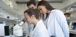
Tu es à la recherche de ton stage de Master ? L’IBDM te semble être le bon endroit pour le faire ? Découvre nos offres dès maintenant.
Félicitation à Robert Kelly, Frank Schnorrer, Cédric Maurange, Bianca Habermann et Delphine Delacour !
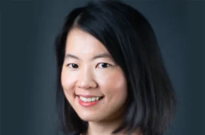
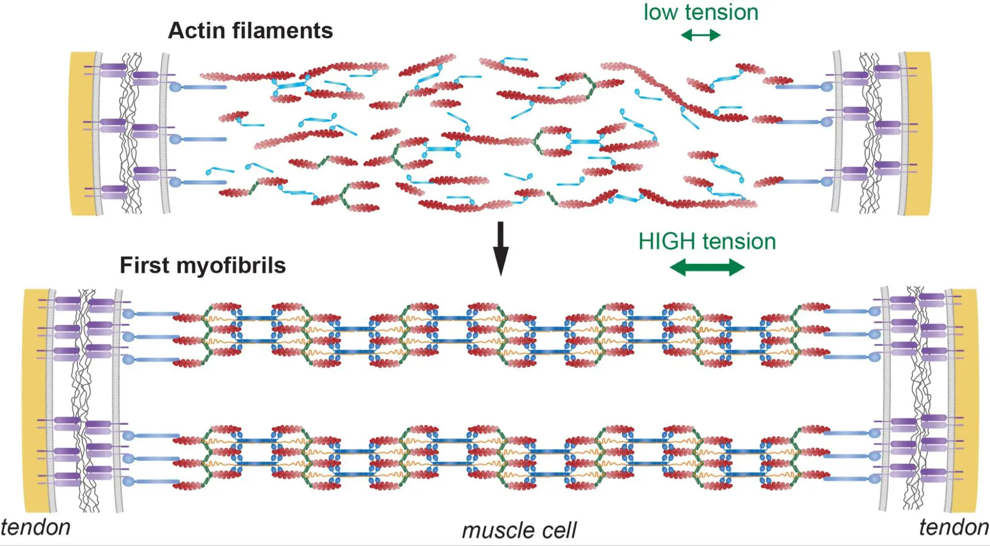
De post-doctorante (bourse AFM-Téléthon) à chercheuse au CNRS : Qiyan Mao fait progresser la recherche sur les muscles humains grâce à des approches en mécanique moléculaire et tissulaire.
L’IBDM inspire les jeunes esprits : en impliquant les enfants des écoles primaires dans la lutte contre le cancer pédiatrique (“Contre le cancer, j’apporte ma pierre”) et en interagissant avec les lycéens grâce à des expériences immersives (DECLICS).

Séminaire Interne par Nuno Luis
Une approche interdisciplinaire pour explorer et cibler la dialectique cancer-cellules immunitaires : d’un modèle de souris TNBC aux patients
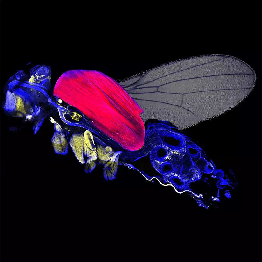
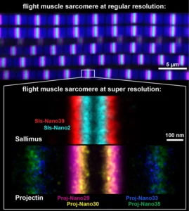
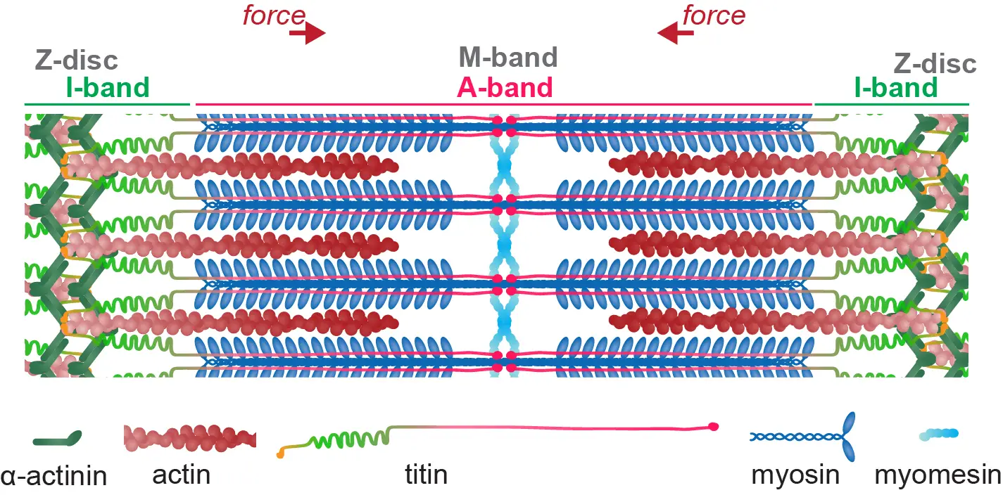
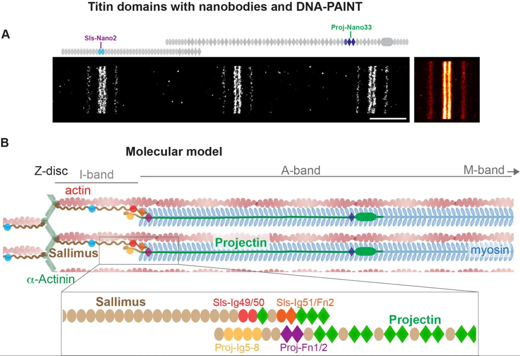
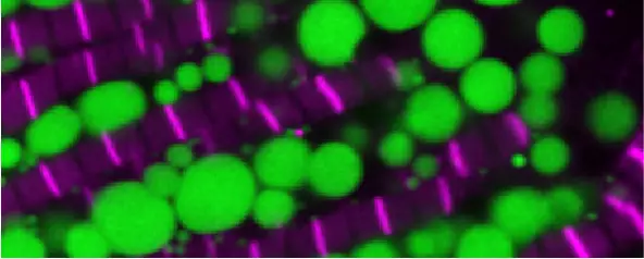
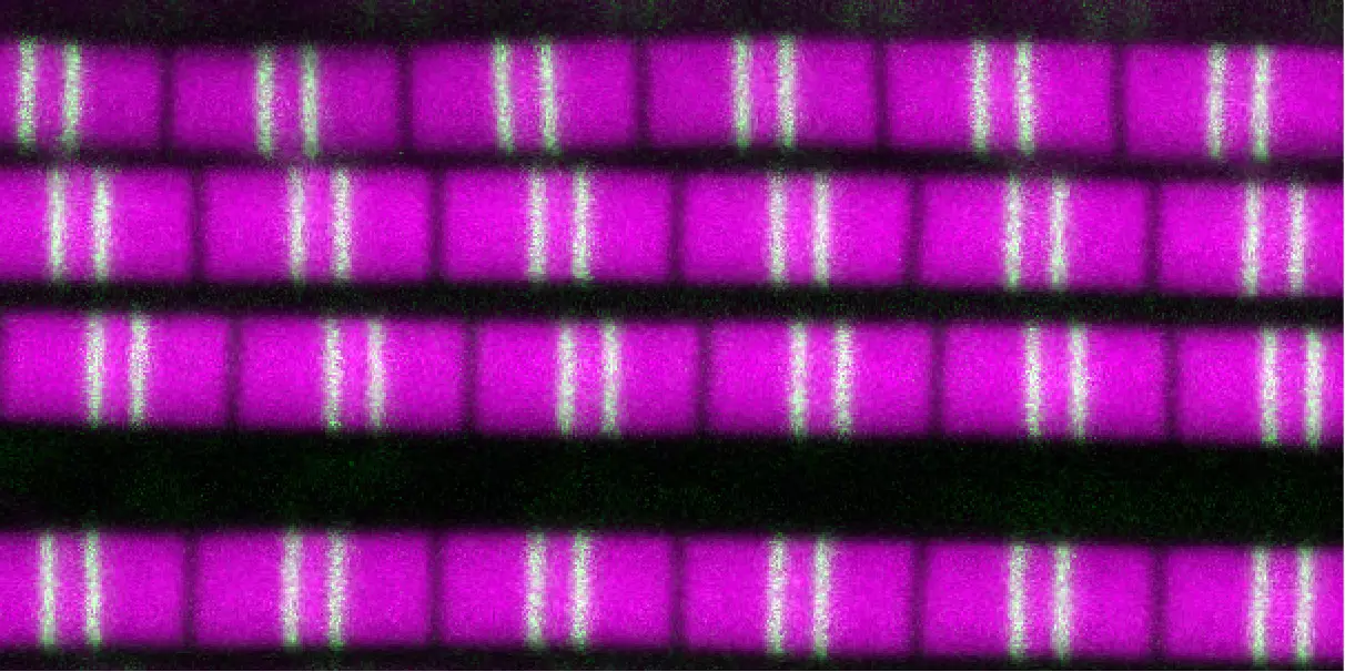
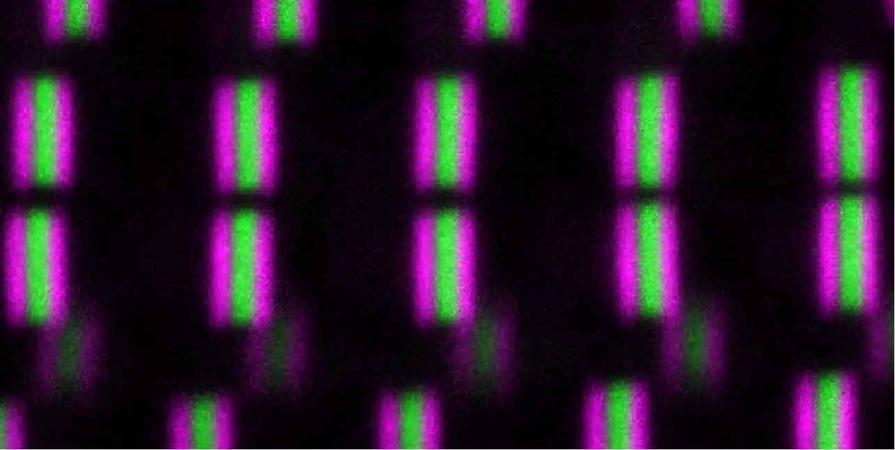
Les sarcomères des drosophiles sondés à l’échelle nanométrique une molécule à la fois à l’aide de nanocorps.
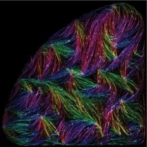
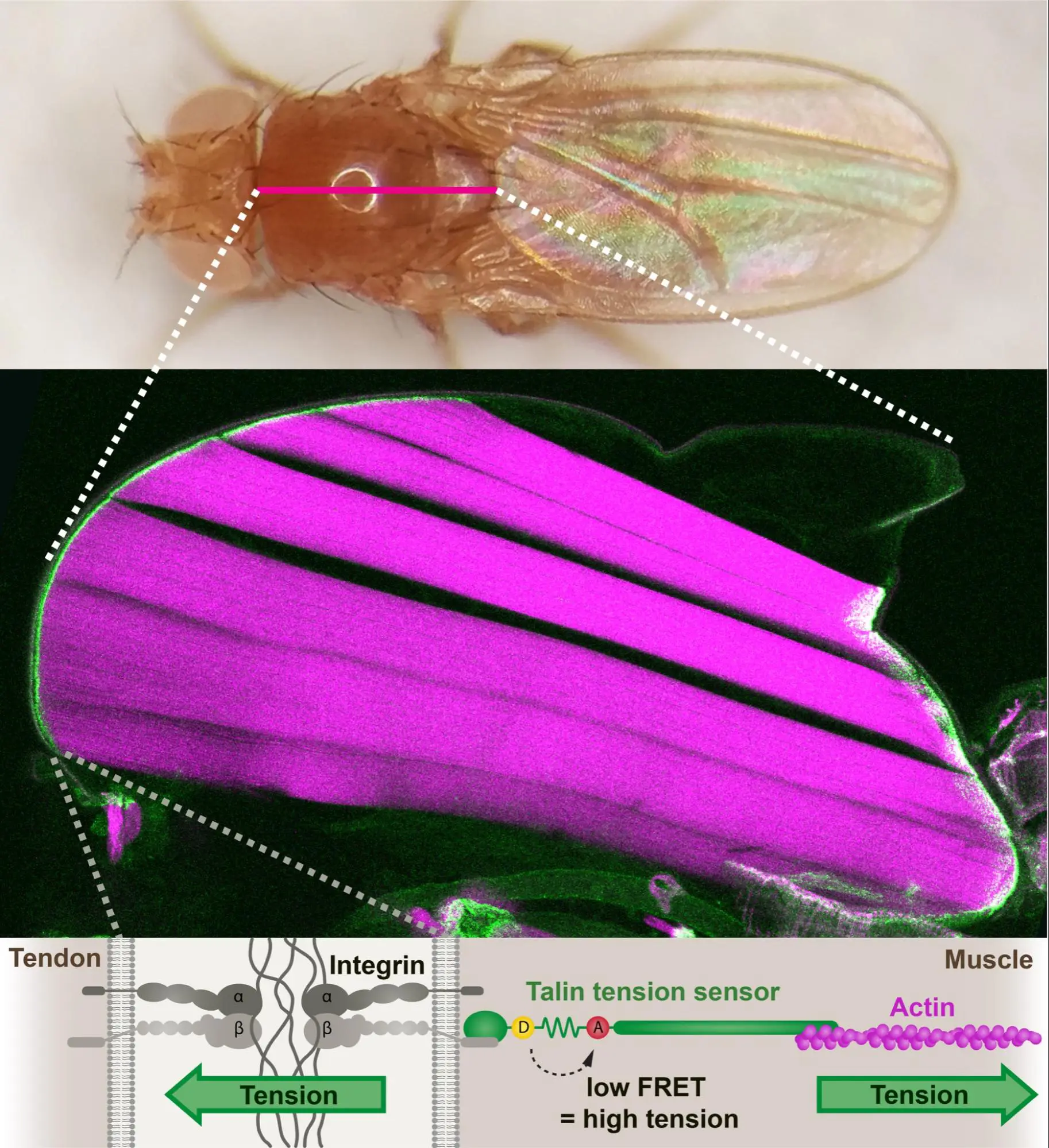
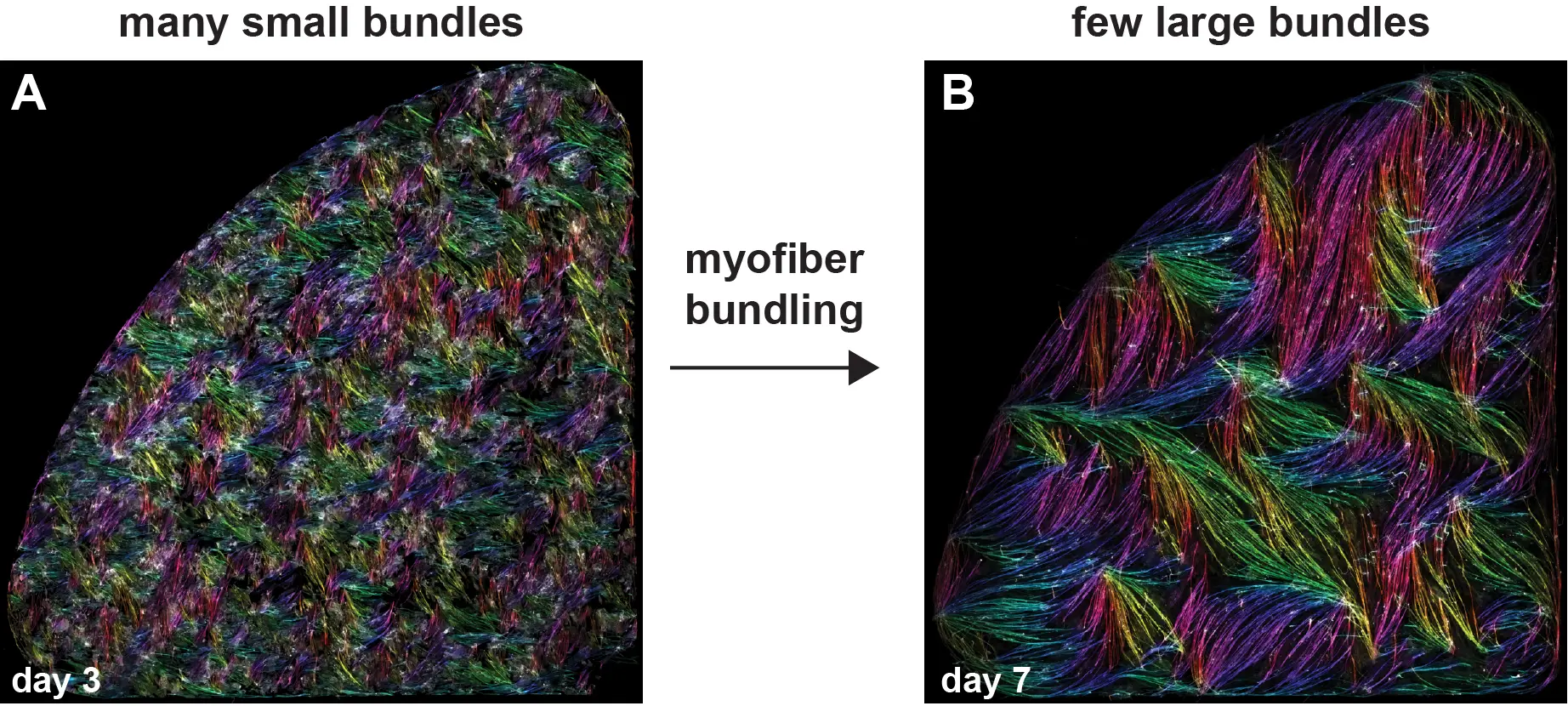
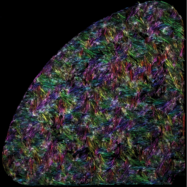
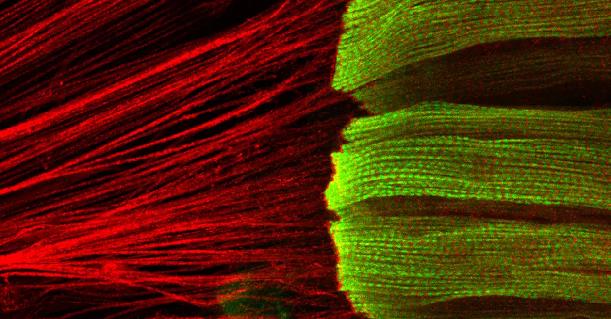
Les cellules musculaires s’auto-organisent en faisceaux de fibres in vitro, sans la présence de signaux externes !
Frank Schnorrer élu membre de l’EMBO
L’EMBO élit 67 nouveaux membres et membres associés. Ils rejoignent la communauté de plus de 1 900 scientifiques du vivant de premier plan en Europe et au-delà.
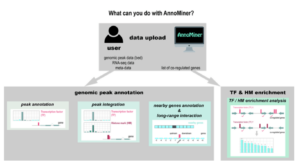
Nous présentons ‘AnnoMiner’, un nouvel outil convivial basé sur le web pour annoter et intégrer les données épigénétiques et de liaison des facteurs de transcription.
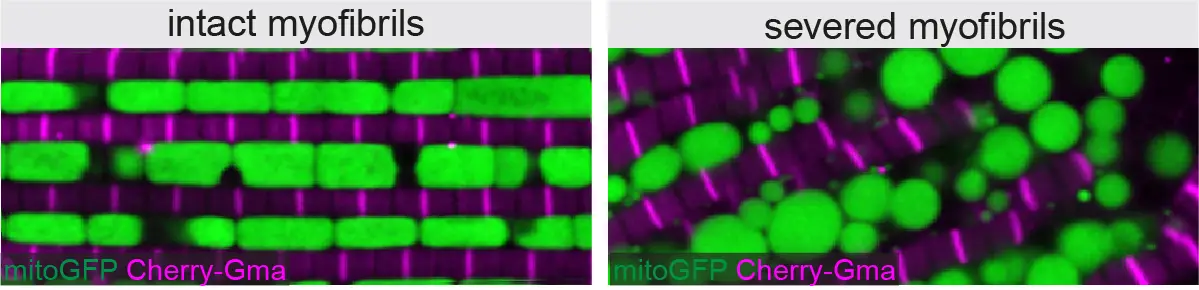
En combinant la génétique chez la drosophile avec l’imagerie de pointe et le “deep-learning”, des chercheurs de l’IBDM ont découvert que les mitochondries coordonnent leur formation avec le développement des myofibrilles pour correspondre au bon type musculaire.
L’équipe de Schnorrer et al. ont découvert qu’une voie de signalisation, appelée voie Hippo, contrôle la croissance musculaire pendant le développement des muscles du vol, chez la drosophile.
Le Conseil européen de la recherche (ERC) a attribué l’un des rares financements ERC Synergy à un consortium international de scientifiques, Frank Schnorrer, Stefan Raunser, Dirk Görlich et Mathias Gautel.
